Home

bölüm liberal aramak cotton wool spots vs hard exudates kriz vergi köpek yavrusu

Classification of Diabetic Retinopathy - YouTube

Two abnormalities: Hard drusen (left) and Cotton wool spots (right). |  Download Scientific Diagram
Two abnormalities: Hard drusen (left) and Cotton wool spots (right). | Download Scientific Diagram

Moran CORE | Retina & RPE Histopathology

Diabetic Retinopathy Flashcards | Quizlet

Cotton-Wool Spots May Challenge Diagnosis

Data on fundus images for vessels segmentation, detection of hypertensive  retinopathy, diabetic retinopathy and papilledema - ScienceDirect
Data on fundus images for vessels segmentation, detection of hypertensive retinopathy, diabetic retinopathy and papilledema - ScienceDirect
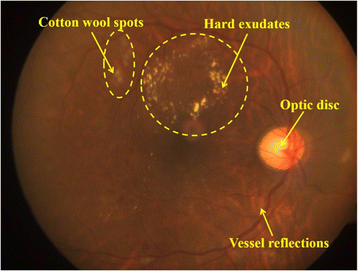
Semi-automated quantification of hard exudates in colour fundus photographs  diagnosed with diabetic retinopathy | BMC Ophthalmology | Full Text
Semi-automated quantification of hard exudates in colour fundus photographs diagnosed with diabetic retinopathy | BMC Ophthalmology | Full Text

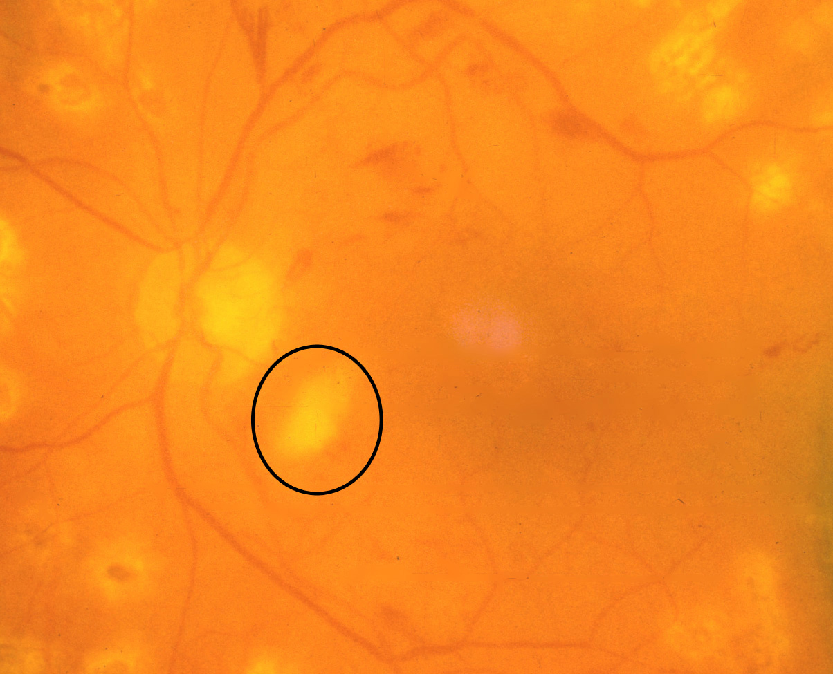
Cotton wool spots. COMS Grading Scheme

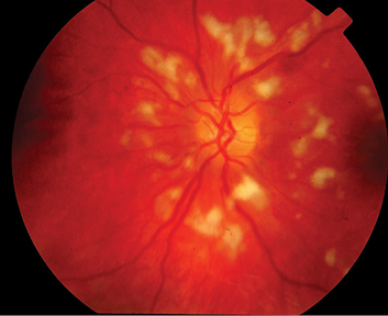
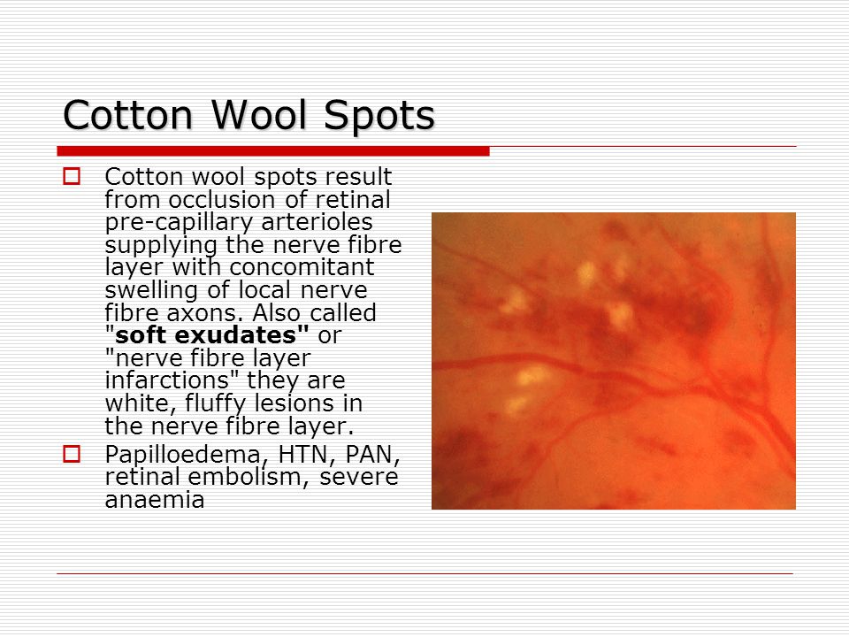
Cotton Wool Spots - EyeWiki

Two abnormalities: Hard drusen (left) and Cotton wool spots (right). |  Download Scientific Diagram
Two abnormalities: Hard drusen (left) and Cotton wool spots (right). | Download Scientific Diagram

A bilateral macular star and optic disc oedema | The BMJ

Diabetic Retinopathy - almostadoctor

Symptoms of retinopathy: (a) hard exudates, (b) cotton wool spots and... |  Download Scientific Diagram
Symptoms of retinopathy: (a) hard exudates, (b) cotton wool spots and... | Download Scientific Diagram

Fundoscopic examination - ppt video online download

Retinal Diseases Flashcards | Chegg.com

Cotton wool spots detection in diabetic retinopathy based on adaptive  thresholding and ant colony optimization coupling support vector machine -  Sreng - 2019 - IEEJ Transactions on Electrical and Electronic Engineering -  Wiley Online Library
Cotton wool spots detection in diabetic retinopathy based on adaptive thresholding and ant colony optimization coupling support vector machine - Sreng - 2019 - IEEJ Transactions on Electrical and Electronic Engineering - Wiley Online Library

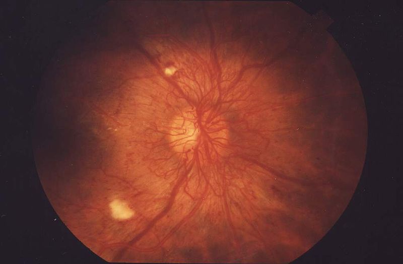
Cotton wool spots - Wikipedia

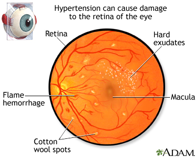
Hypertensive retinopathy - Wikipedia

Diabetic Retinopathy Non Proliferative Illustration Shows Hard Exudates  Cotton Wool Stock Photo by ©katerynakon 567244272
Diabetic Retinopathy Non Proliferative Illustration Shows Hard Exudates Cotton Wool Stock Photo by ©katerynakon 567244272

Hypertensive retinopathy: MedlinePlus Medical Encyclopedia Image

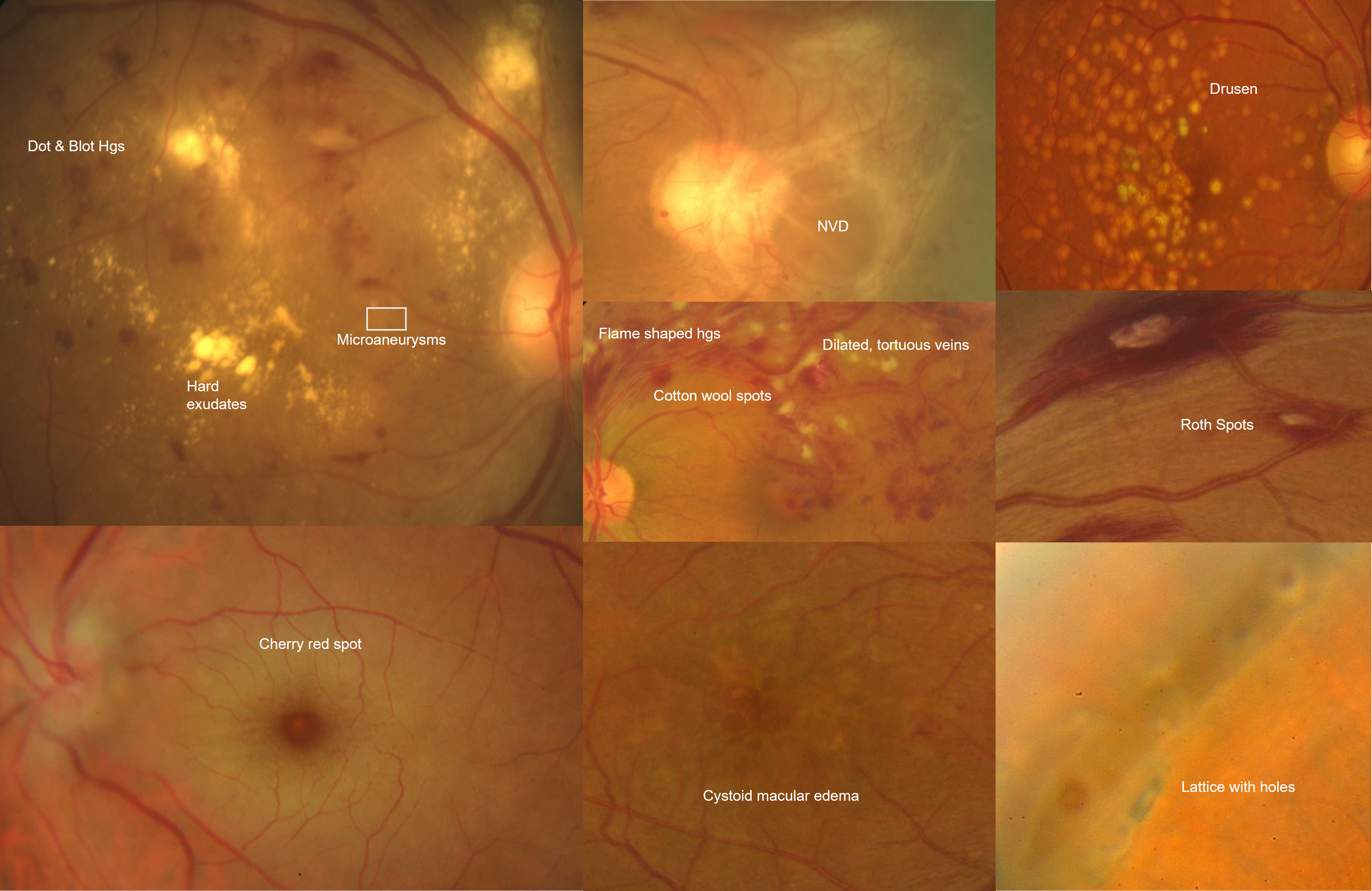
Ophthalmology-Notes And Synopses - Layers of Retina affected in Diabetic  Retinopathy: ➖Cotton Wool Spots: Nerve fibre layer. ➖Microaneursyms: Inner  nuclear layer. ➖Dot blot hemorrhages: Inner nuclear & Outer plexiform  layer. ➖Flame-shaped hemorrhages:
Ophthalmology-Notes And Synopses - Layers of Retina affected in Diabetic Retinopathy: ➖Cotton Wool Spots: Nerve fibre layer. ➖Microaneursyms: Inner nuclear layer. ➖Dot blot hemorrhages: Inner nuclear & Outer plexiform layer. ➖Flame-shaped hemorrhages:

PDF] Detection Of Cotton Wool Spots In Retinopathy Images : A Review |  Semantic Scholar
PDF] Detection Of Cotton Wool Spots In Retinopathy Images : A Review | Semantic Scholar

Figure 1 from Automated detection and differentiation of drusen, exudates,  and cotton-wool spots in digital color fundus photographs for diabetic  retinopathy diagnosis. | Semantic Scholar
Figure 1 from Automated detection and differentiation of drusen, exudates, and cotton-wool spots in digital color fundus photographs for diabetic retinopathy diagnosis. | Semantic Scholar

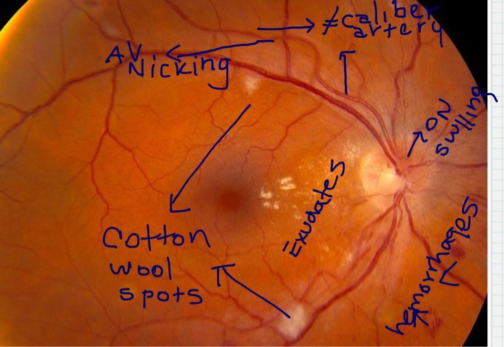
Ten Clinical Signs A Postgraduate Must Look For While Examining the Fundus